Overview: What Kind of Test Is This?
The ABI test is a non-invasive vascular study. "Non-invasive" means nothing goes into your body — no needles, no injections, no contrast dye, no radiation. You simply lie on an examination table while a technician takes blood pressure readings at your arms and ankles using a blood pressure cuff and a small hand-held device.
The test is completely painless for the vast majority of patients. The only sensation you'll feel is the familiar squeeze of a blood pressure cuff inflating and then releasing — the same thing you experience at every regular doctor's visit. The entire procedure typically takes 10 to 20 minutes for a complete bilateral study (both legs).
Duration: 10–20 minutes | Equipment: Blood pressure cuff + Doppler probe | Invasiveness: None | Radiation: None | Cost: $150–$400 out-of-pocket, often covered by insurance when medically necessary
Before Your Test: Preparation
The ABI test requires very little preparation, which is one of its great advantages. Here's what you should know:
- No fasting required. You can eat and drink normally before the test.
- Rest for 10–30 minutes before the test if you've been active. Walking increases blood flow and can temporarily elevate ankle pressure readings. Most vascular labs will have you lie down and rest for 5–10 minutes in the lab before measuring begins.
- Wear comfortable, loose-fitting clothes — you'll need to expose your upper arms and the area just above your ankles. Socks and shoes will need to come off.
- Tell your technician about any medications you're taking, especially blood pressure medications. These don't need to be stopped — they're part of your normal baseline — but the technician should know about them.
- Avoid smoking for at least one hour before the test. Nicotine causes temporary arterial constriction that can artificially lower your ABI reading.
- If you have open wounds or active ulcers on your legs or feet, tell your doctor before scheduling. The test can still be done, but the technician needs to take precautions.
Equipment Used for an ABI Test
You'll encounter just two pieces of equipment during the test:
1. Blood Pressure Cuffs
Standard blood pressure cuffs — the kind used for routine checks at every doctor's office — are used for both the arm and ankle measurements. The cuffs for the ankle are the same width as arm cuffs but placed above the ankle. The cuff inflates to temporarily cut off blood flow, then slowly deflates while the technician listens for the return of flow.
2. Hand-Held Doppler Ultrasound Probe
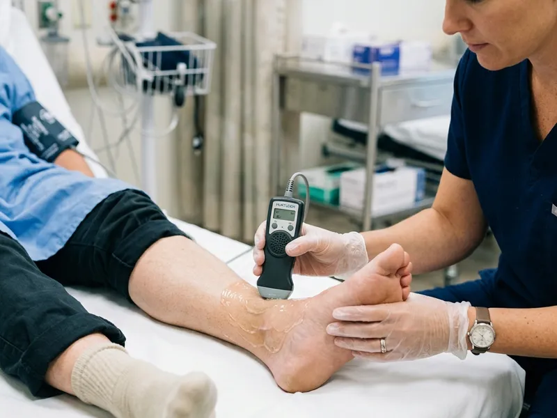
This is the key tool that makes the ABI different from a standard blood pressure check. A regular stethoscope can't reliably detect the faint pulse signals in ankle arteries. The Doppler probe — which looks like a small pen or wand — uses ultrasound waves to detect moving red blood cells. When blood starts flowing again as the cuff deflates, the Doppler picks up the characteristic "whooshing" or "rushing" sound and indicates the pressure at which flow returns.
The Doppler probe is simply held against the skin over the ankle artery with a small amount of gel (the same type used for an ultrasound scan). It makes no contact with any broken skin and causes no discomfort.
The ABI Test: Step by Step
Here's exactly what happens during a standard bilateral (both legs) ABI test:
Step 1 — Arrive and Lie Down (0–10 min)
You'll be taken to a quiet room and asked to lie flat on an examination table. You'll be given 5 to 10 minutes to rest, allowing your blood pressure to stabilize from any walking or activity. Remove your socks and shoes before resting.
Step 2 — Arm Measurements (2–4 min)
The technician places blood pressure cuffs on both of your upper arms and takes a reading on each side. A standard automated cuff or manual sphygmomanometer is used for this step — no Doppler needed here. The highest reading from either arm is recorded as the denominator in your ABI calculation.
Step 3 — Ankle Measurements (5–10 min)
Cuffs are placed just above each ankle. The technician applies a small amount of gel over the ankle artery location and places the Doppler probe against the skin. The cuff inflates until the Doppler signal disappears (meaning flow is cut off), then slowly deflates. The pressure at which the Doppler signal returns is recorded as the ankle systolic pressure. This is done at two artery sites on each ankle: the posterior tibial artery (behind the inner ankle bone) and the dorsalis pedis artery (on the top of the foot). The higher of the two ankle values on each side is used.
Step 4 — Calculation (1 min)
The ABI for each leg is calculated: highest ankle pressure ÷ highest arm pressure. This gives you two numbers — one for the right leg, one for the left. The lower of the two is typically the clinically significant finding.
Step 5 — Results Discussion (2–5 min)
Your results may be discussed with you immediately by the technician or vascular nurse, or they may be reported to your referring physician who will contact you separately. Either way, you should receive your ABI numbers and a brief interpretation of what they mean.
Exercise ABI Testing — When Rest ABI Isn't Enough
Some patients experience leg pain and poor circulation only during physical activity — but their resting ABI comes back normal. This scenario is more common than most people realize. For these patients, an exercise ABI test provides the missing picture.
The protocol typically involves:
- Recording resting ABI values as normal
- Having the patient walk on a treadmill (typically at a standardized speed and incline, such as 2 mph at 12% grade) until they experience their usual symptoms or reach a maximum time limit (usually 5 minutes)
- Immediately after stopping, ankle pressures are remeasured at 1, 3, and 5 minutes of recovery
- A significant drop in ankle pressure after exercise (greater than 20 mmHg) compared to the pre-exercise value is diagnostic for exercise-induced PAD, even when the resting ABI is normal
If you have classic claudication symptoms (leg pain when walking that stops with rest) but a normal resting ABI, ask your doctor specifically about an exercise ABI test. It can be the difference between a diagnosis and being told "everything looks fine."
Understanding and Acting on Your Results
You'll receive two numbers: your right leg ABI and your left leg ABI. Reference these against the standard ranges (detailed in our ABI normal range guide). A few points to keep in mind:
- The lower of the two ABI values is the one that drives clinical decisions
- If only one leg has a low ABI, the disease may be localized to that side — which actually helps guide treatment planning
- A single ABI test is a snapshot in time. Your doctor may repeat it in 6 months or a year to track trends
- ABI results should always be interpreted in the context of your symptoms. A borderline ABI with classic claudication is clinically more significant than a borderline ABI with no symptoms at all
Who Performs the ABI Test?
ABI tests can be performed in several settings across the USA and Canada:
- Vascular surgery clinics and vascular labs — the most common setting; technicians are registered vascular technologists (RVT) certified by the Alliance for Vascular Access Teaching and Research (AVASTAR) or the American Registry of Diagnostic Medical Sonography (ARDMS)
- Cardiology practices — particularly those with non-invasive vascular labs
- Primary care offices — some primary care physicians perform basic ABI screening in their offices, particularly for high-risk patients
- Wound care centers — always measure ABI before applying compression therapy
- Hospital outpatient departments
In Canada, ABI tests are typically ordered by a physician (family doctor, internist, or specialist) and performed at a hospital vascular lab or at a community diagnostic imaging center. Coverage under provincial health insurance plans varies — see our guide to PAD resources in Canada for province-specific information.
For information about what the test costs and how to get insurance coverage, see our dedicated ABI test cost and insurance guide.