If you have diabetes and your ABI reading is above 1.40 — this does not mean excellent arterial health. It almost always means your arteries are too stiff and calcified for the ABI test to give a reliable result. Ask your doctor about a Toe-Brachial Index (TBI) test instead.
Why Diabetes and ABI Don't Always Mix Well
The ABI test works by using a blood pressure cuff to temporarily compress the artery until blood flow stops. When the cuff slowly deflates, the pressure at which blood flow returns is recorded. For this to work accurately, the artery must be compressible — meaning it needs some degree of elasticity and flexibility.
Here's the problem with diabetes: long-standing high blood glucose levels cause a specific type of arterial damage called medial arterial calcification (MAC) — the deposition of calcium crystals in the middle layer (tunica media) of the artery wall. Unlike the plaque that builds up on the inner surface of the artery in atherosclerosis, medial calcification makes the artery wall itself rigid and pipe-like.
A calcified artery simply cannot be compressed by the cuff. So the cuff inflates, inflates, inflates — and the Doppler probe keeps picking up blood flow because the stiff wall won't collapse. The recorded pressure is artificially high — sometimes exceeding 200–300 mmHg in severely calcified arteries, giving an ABI of 1.5, 1.8, or even higher. This is not healthy; it's an artifact of the test's limitation.
The Prevalence of Medial Calcification in Diabetes
How common is this problem? Studies using plain X-rays to detect visible arterial calcification have found:
- Approximately 30–40% of diabetic patients with PAD have calcification severe enough to make the ABI unreliable (ABI >1.40)
- The risk increases with diabetes duration — patients with 10+ years of diabetes are at highest risk
- Patients with diabetic kidney disease (nephropathy) or end-stage renal disease on dialysis have particularly severe MAC — some studies find unreliable ABI in >60% of dialysis-dependent diabetics
- Poor glycemic control accelerates MAC progression — each percentage point increase in HbA1c above target correlates with measurably faster calcification
Diabetic PAD: A More Dangerous Form of the Disease
Beyond the testing challenges, PAD in diabetic patients has several features that make it more dangerous than PAD in non-diabetics:
1. More Distal Disease
Non-diabetic PAD tends to preferentially affect the larger, proximal arteries — the aorta, iliac arteries, and femoral arteries. Diabetic PAD characteristically affects the more distal (below-the-knee) arteries — the tibial and peroneal arteries, and often the foot arteries. This distal distribution is clinically important because:
- Distal disease is technically more challenging to treat with endovascular procedures
- Distal disease means the foot — the most vulnerable part — has the least blood flow
- Diabetic foot ulcers develop in a context of both arterial insufficiency (PAD) and neuropathy, creating a particularly dangerous combination
2. Peripheral Neuropathy Masks Symptoms
Up to 50% of long-standing diabetics have peripheral neuropathy — nerve damage that reduces or eliminates sensation in the feet and lower legs. This has a critical implication: a diabetic patient can have severe PAD, non-healing foot ulcers, and even impending tissue death — without feeling significant pain. The warning signal that would drive a non-diabetic to the emergency room is simply absent.
This is one of the primary reasons why diabetic foot complications can progress so rapidly and why regular vascular screening (even when asymptomatic) is critical in this population.
3. Higher Infection Risk
Diabetes impairs immune function and healing. When a diabetic patient has a foot wound and inadequate blood flow, healing is doubly compromised: the blood isn't delivering sufficient oxygen and nutrients, AND the immune response is blunted. Minor infections can become limb-threatening cellulitis or osteomyelitis (bone infection) within days.
4. Worse Outcomes After Revascularization
Diabetic patients who undergo endovascular or surgical revascularization for PAD have higher rates of restenosis (the artery re-narrowing), wound complications, and amputation compared to non-diabetics — though outcomes have improved significantly with contemporary techniques and better glucose management.
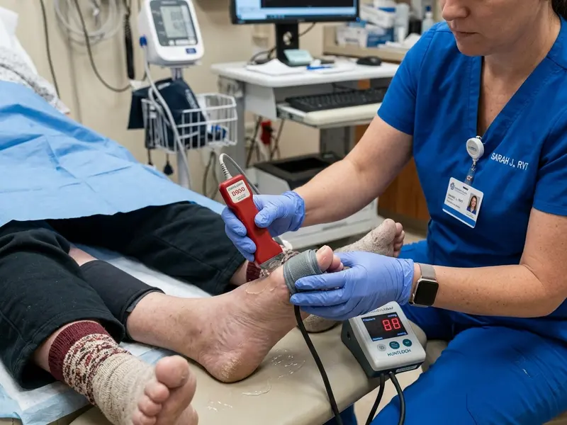
The Toe-Brachial Index (TBI): The Solution for Diabetics
The Toe-Brachial Index (TBI) solves the medial calcification problem elegantly. The digital arteries in the toes are far smaller in diameter and have very little smooth muscle in their walls — they simply don't develop the same calcification that affects larger leg arteries. As a result, they remain compressible even in patients with severe medial calcification elsewhere.
The TBI uses a tiny blood pressure cuff placed around the base of the big toe, and a highly sensitive photoplethysmography (PPG) sensor (rather than a Doppler probe) to detect the return of blood flow as the cuff deflates. This technique accurately measures toe systolic pressure, which can then be compared to arm systolic pressure to give the TBI ratio.
| TBI Score | Classification | Clinical Significance |
|---|---|---|
| > 0.70 | Normal | Adequate toe perfusion; no significant PAD |
| 0.60 – 0.70 | Borderline | Early PAD; monitor closely |
| 0.40 – 0.59 | Mild-Moderate PAD | Significant arterial disease; treatment indicated |
| < 0.40 | Severe PAD / Possible CLTI | Urgent evaluation needed |
| Absolute toe pressure <30 mmHg | CLTI | Critical limb-threatening ischemia; urgent revascularization |
Note that TBI normal reference ranges are lower than ABI ranges, because toe systolic pressures are normally slightly lower than ankle pressures even in healthy individuals.
ABI Screening Guidelines for Diabetic Patients
The American Diabetes Association (ADA) and the ACC/AHA both recommend structured vascular assessment for diabetic patients:
- ABI (or TBI if ABI >1.4) should be performed in all diabetic patients with any symptoms of PAD
- Consider ABI screening in all diabetic patients over age 50, even without symptoms
- In diabetic patients with foot problems (non-healing wounds, infections, or amputations), vascular assessment is mandatory before wound management
- The ADA's Standards of Medical Care in Diabetes (2026) recommend annual foot examination for all diabetic patients — which should include checking for diminished pulses and signs of ischemia
Practical PAD Management for Diabetic Patients
If you have diabetes and are concerned about PAD, here's a practical framework:
- Get an ABI test annually from your podiatrist, internist, or vascular specialist — even if you have no symptoms. If ABI >1.4, request a TBI.
- Inspect your feet daily. Look for blisters, cuts, redness, swelling, or discoloration. Use a mirror to see the sole. Report any wound that hasn't healed in 2 weeks immediately.
- Never walk barefoot — even indoors. Diabetic neuropathy means you may not feel a cut or blister until significant damage has occurred.
- Choose proper footwear. Poorly fitting shoes are a leading cause of diabetic foot ulcers. Consider custom orthotics or therapeutic footwear — Medicare covers therapeutic shoes for qualifying diabetics.
- Control your blood sugar aggressively. HbA1c target below 7% (ideally 6.5%) slows both neuropathy and PAD progression. GLP-1 agonists (semaglutide, liraglutide) and SGLT-2 inhibitors have additional cardiovascular benefits beyond glucose control and should be discussed with your endocrinologist.
- Work with a multidisciplinary team: Optimal diabetic PAD management requires collaboration between your primary care physician, endocrinologist, vascular specialist, podiatrist, and when needed, a wound care nurse.